Scalp dermoscopy is a technique that has been recently utilized for the diagnosis, follow-up, treatment and prognosis of hair and scalp disorders. It presents a valuable link between clinical features and histological diagnosis (biopsy). It is also a way to avoid unnecessary biopsies.
For scalp examination, there is manual dermoscope or videodermoscope that has higher magnification.
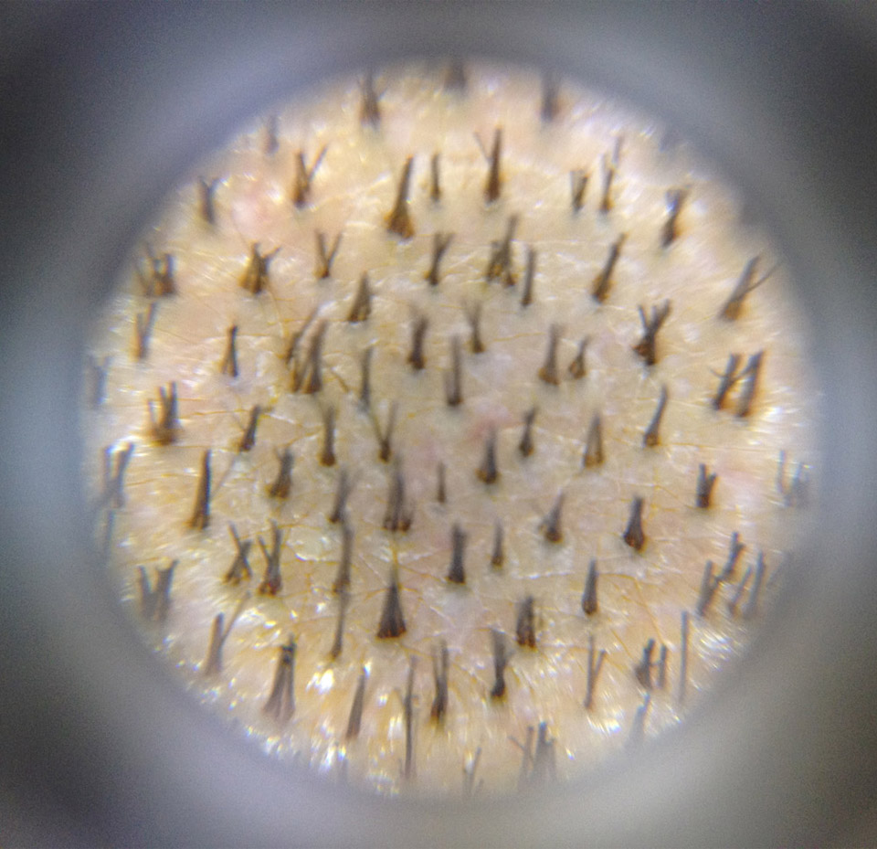
This permits evaluation of:
- scalp condition;
- follicular and perifollicular abnormalities
- hair shaft characteristics
- hair density.
Scalp condition is very important to be examined for, compared to what are the hallmarks of a healthy scalp. Trichoscopy can reveal any hypo or hyperpigmentation of the scalp. Scaly conditions such as seborrheic dermatitis and psoriasis can be differentiated with trichoscopy. Also, cutaneous microvasculature is visualized.
Hair shaft characteristics are visualized with dermoscopy and changes are clearly defined, such as the difference between normal (terminal) hair and vellus hair, exclamation hairs in alopecia areata, as well as other hair shaft abnormalities and diameter differences.
Hair density is crucial in diagnosing certain hair conditions, along with the ratio of vellus hair to terminal hair-the grade of miniaturization. This is all easily evaluated with dermoscopy and it is called densitometry.
Trichoscopy may help to distinguish scarring versus non-scarring alopecia, early androgenetic alopecia (AGA) versus telogen effluvium (TE), and it also supports the diagnosis and predicts the prognosis of alopecia areata (AA).
Typical areas of the scalp for dermoscopy are frontal, occipital and parietal. If hair loss is localized, then that certain area is examined. The area is first immersed with alcohol and then the evaluation is performed. Several pictures are taken and evaluated. This is a precious tool to compare different treatments of hair loss.
